Testing Department
The American Eye and Retina Canter has the latest advanced eye examination equipment.
Tomographic examination of the cornea with the device of the Pentacam
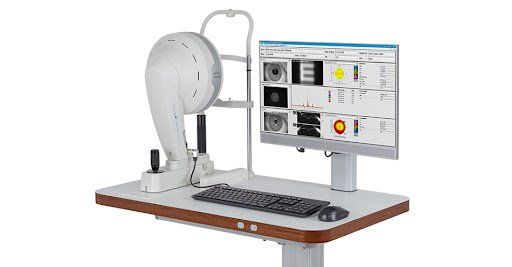
A stratified image of the cornea with the PentaCam device: This examination takes a stratified image of the cornea of the eye. This image helps to know the health of the cornea from its shape and thickness. This device also gives very accurate information that helps in choosing the type of vision correction operation. The examinations of this device can show the stages of keratoconus disease and the disease can be diagnosed in its early stages and treated early.
A stratified image of the cornea with the PentaCam device: This examination takes a stratified image of the cornea of the eye. This image helps to know the health of the cornea from its shape and thickness. This device also gives very accurate information that helps in choosing the type of vision correction operation. The examinations of this device can show the stages of keratoconus disease and the disease can be diagnosed in its early stages and treated early.
Measuring the power of the lens with the IOL Master
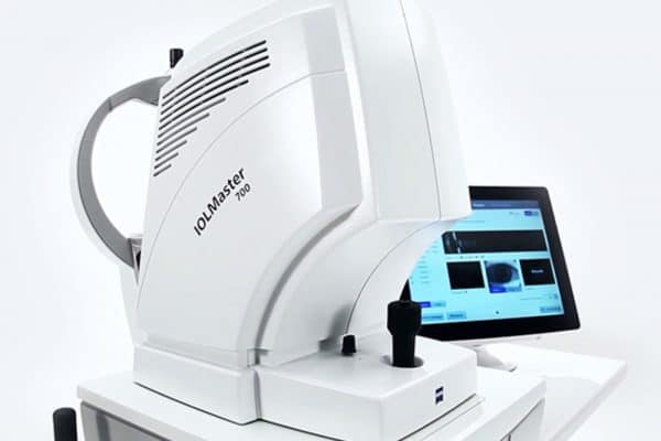
Measuring the lens of the eye with the IOL Master device. The American Centre for Eye and Retina has the latest device to measure the lens of the eye. This device is used to determine the strength of the lens that must be implanted to the patient during the cataract operation. Therefore, this device is one of the important and accurate devices used in cataract operations and has a role Great in the calculations of the specialist for the implanted lens, on which the results of the operation depend on the clarity and accuracy of vision.
Measuring the lens of the eye with the IOL Master device. The American Centre for Eye and Retina has the latest device to measure the lens of the eye. This device is used to determine the strength of the lens that must be implanted to the patient during the cataract operation. Therefore, this device is one of the important and accurate devices used in cataract operations and has a role Great in the calculations of the specialist for the implanted lens, on which the results of the operation depend on the clarity and accuracy of vision.
Retinal examination with Fundus Camera
The colour image of the retina with the Fundus Camera. This device performs a careful and detailed examination of all the minute arteries in the retina. This image helps identify the damaged areas in the retina and determine the areas of bleeding, if any, which helps the specialist doctor to treat the condition accurately.
The colour image of the retina with the Fundus Camera. This device performs a careful and detailed examination of all the minute arteries in the retina. This image helps identify the damaged areas in the retina and determine the areas of bleeding, if any, which helps the specialist doctor to treat the condition accurately.
Tomographic examination of the retina and optic nerve using the OCT
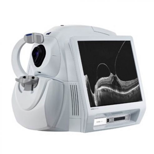
A tomographic image of the retina and optic nerve with the OCT device. This device examines a tomographic image of the retina, showing the layers of the retina and clarifying which layer of the retina is damaged, as it shows if there is a fluid accumulation under the retina. This device also performs a tomography of the optic nerve and thus shows the health of the optic nerve and shows the damage The optic nerve, which often results from high eye pressure, glaucoma, or some genetic or other diseases. All this accurate information from this device helps the specialist doctor quickly and accurately diagnose the patient’s condition and treat it in the right way, knowing that the American Eye and Retina device is one of the latest The devices in the world and the most accurate, which is of high quality and accuracy in the examination.
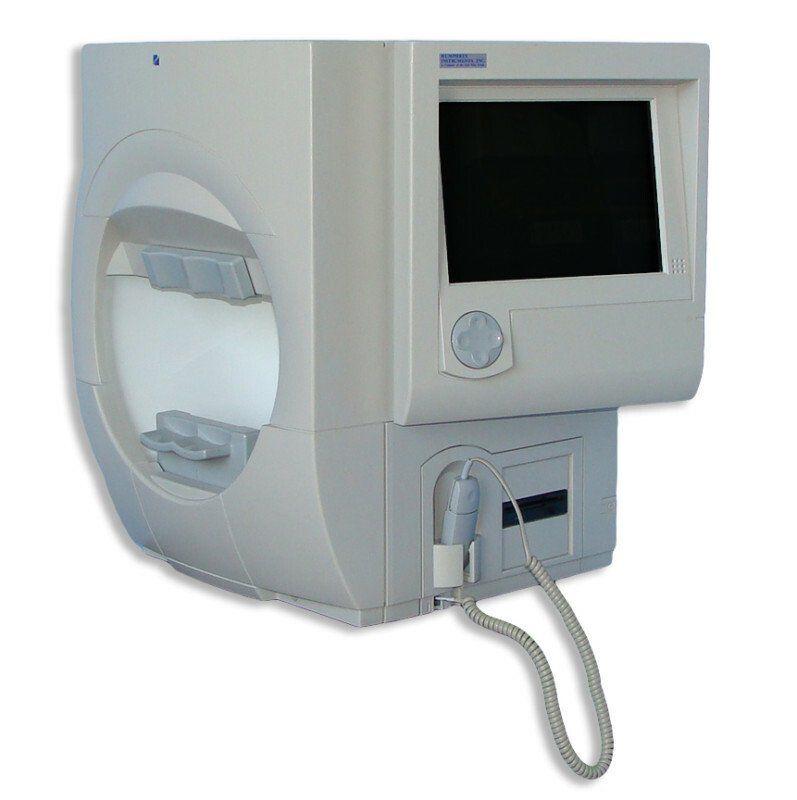
Examination of the visual field with a visual field device
Examination of the visual field with the Visual Field device. This examination is one of the most important types of examinations that patients with high intraocular pressure, glaucoma, and glaucoma need. This device helps determine the area through which the patient can see through. With this examination, the percentage of damage in the optic nerve and the stage the patient has reached can be diagnosed. It helps in treating the patient and following up on the condition.
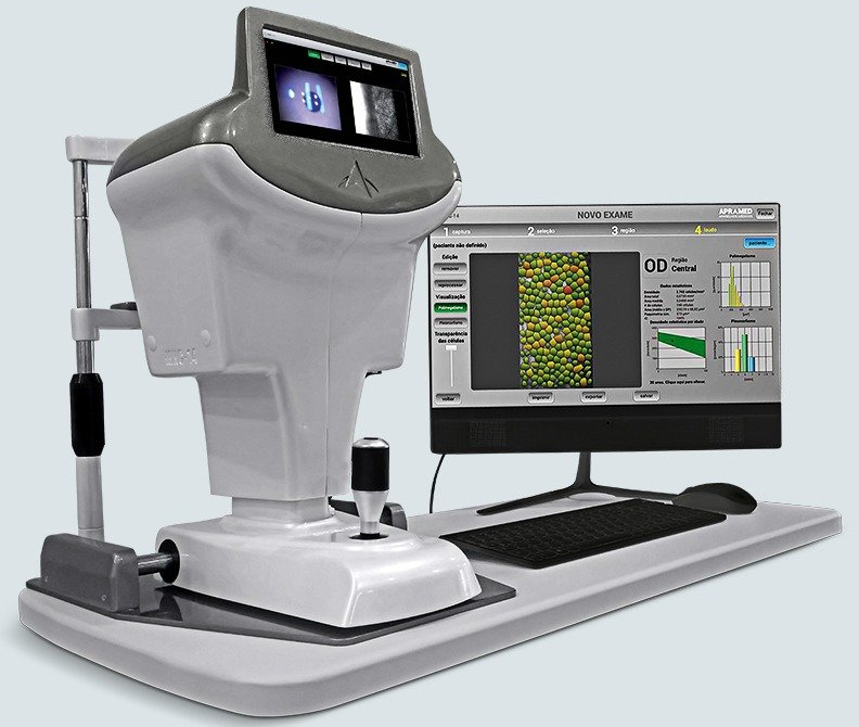
Examination of the inner cells of the cornea with a Specular Microscope
The American Centre for the Eye and Retina has the most important and latest devices in the world in ophthalmology and surgery. This device examines the number of cells in the inner layer of the cornea, the endothelial cell. This examination is one of the most important tests used in cataract operations and corneal transplants. Through this technology, the specialist doctor can approximate the success rate of these operations, choose the appropriate operation for the patient, and give treatments that help after the operation. With this device, which the American Eye and Retina Centre has provided, it has helped a large number of patients to obtain more accurate and better results in their operations.
The American Centre for the Eye and Retina has the most important and latest devices in the world in ophthalmology and surgery. This device examines the number of cells in the inner layer of the cornea, the endothelial cell. This examination is one of the most important tests used in cataract operations and corneal transplants. Through this technology, the specialist doctor can approximate the success rate of these operations, choose the appropriate operation for the patient, and give treatments that help after the operation. With this device, which the American Eye and Retina Centre has provided, it has helped a large number of patients to obtain more accurate and better results in their operations.


